Israeli scientists created the world’s first 3D-printed heart using human cells

Happy Memorial Day Holiday! As coronavirus overwhelms the health care system, people with other illnesses are struggling to find treatment. The fight against coronavirus has shifted attention away from patients suffering from cardiovascular disease and other chronic and life threatening diseases.
According to CDC, one person dies every 37 seconds in the United States from cardiovascular disease. About 647,000 Americans die from heart disease each year—that’s 1 in every 4 deaths. That’s more than the number of people who died from the ongoing coronavirus pandemic. Heart disease costs the United States about $219 billion each year from 2014 to 2015. Cardiovascular diseases are the number one cause of death in industrialized nations. To date, heart transplantation is the only treatment for patients with end‐stage heart failure. Since the number of cardiac donors is limited, there is a need to develop new approaches to regenerate the infarcted heart.
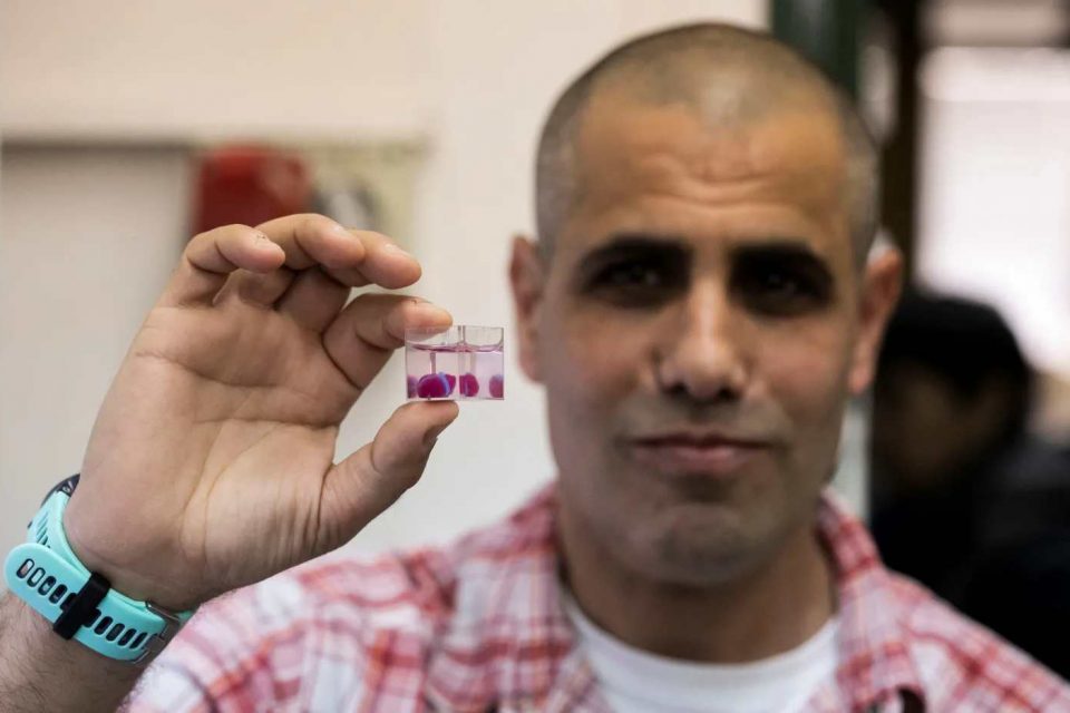
However, this is hope. Scientists at Tel Aviv University in Israel have printed the world’s first 3D heart complete with blood vessels using a patient’s tissue, vessels, collagen, and biological molecules. Even though challenges remain, The extraordinary breakthrough could one day be used to heal hearts or engineer new ones for transplants and render organ donation obsolete.
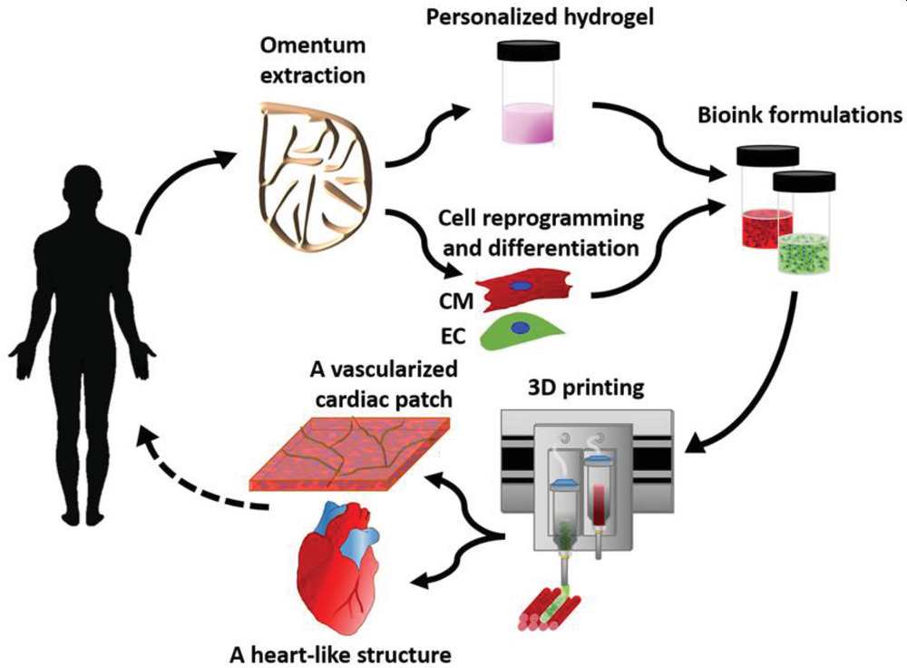
To “print” the 3D printed heart, Israeli scientists used “personalized hydrogel” made of collagen, a protein that supports the cell structures, to form “bioinks.” The hydrogel originated from fatty tissues extracted from human test subjects. The breakthrough was reported by lead scientists Prof. Tal Dvir, Dr. Assaf Shapira of TAU’s Faculty of Life Sciences and Nadav Noor, his doctoral student, in Advanced Science journal.
Among the challenges that remain is finding a way to generate enough cells to create the tissue required to “print” a human-sized heart. True, the heart is the size of a rabbit’s, and it doesn’t actually work yet. Dvir pointed out however that “printing” a human-size heart involves basically the same technology.
Below is an excerpt of how the scientists described the process:
Here, a simple approach to 3D‐print thick, vascularized, and perfusable cardiac patches that completely match the immunological, cellular, biochemical, and anatomical properties of the patient is reported. To this end, a biopsy of an omental tissue is taken from patients. While the cells are reprogrammed to become pluripotent stem cells, and differentiated to cardiomyocytes and endothelial cells, the extracellular matrix is processed into a personalized hydrogel. Following, the two cell types are separately combined with hydrogels to form bioinks for the parenchymal cardiac tissue and blood vessels. The ability to print functional vascularized patches according to the patient’s anatomy is demonstrated.

By the way, this is not the first time 3D printing technology was used to print organs. However, this is the first time human cells are used to print organs. For example, in 2017, a team of researchers at ETH Zurich created a 3D printed artificial heart. But rather than using human tissue, the researchers used a flexible material.
Below are two videos of how their breakthrough.

