Scientists at Carnegie Mellon advances FRESH 3D bioprinting to rebuild functional part of human heart, using a technology developed by startup FluidForm

Scientists have taken a major step closer to being able to 3D bioprint functional organs, after researchers devised a novel method of rebuilding components of the human heart, according to a study published in the August 2nd edition of Science.
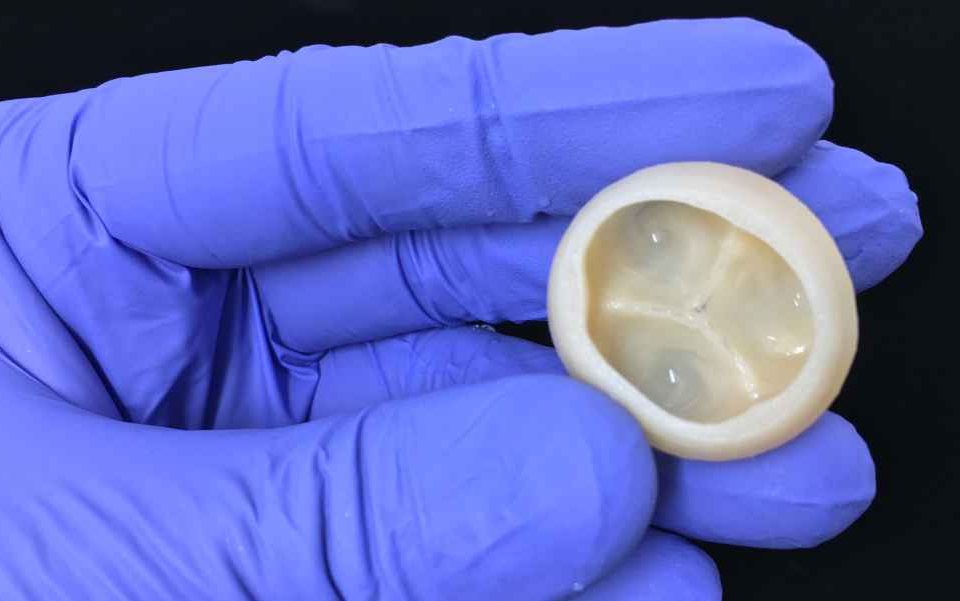
The scientists from Carnegie Mellon University (CMU), successfully developed an advanced version of Freeform Reversible Embedding of Suspended Hydrogels (FRESH) technology to 3D print collagen for small blood vessels, valves, and beating ventricles. The new novel 3D bioprinting method is used to build functional parts of the human heart.
FRESH technology is patented to FluidForm, a Massachusetts-based medical startup. The technology was recently awarded US patent 10,150,258, FRESH™ technology is now licensed to FluidForm, a startup committed to dramatically expanding the capability of 3D printing.
“We now have the ability to build constructs that recapitulate key structural, mechanical, and biological properties of native tissues,” said Prof. Adam Feinberg, CTO and co-founder, FluidForm, and Principal Investigator, Regenerative Biomaterials and Therapeutics Group, Carnegie Mellon, where the research was done. “There are still many challenges to overcome to get us to bioengineered 3D organs, but this research represents a major step forward.”
Though 3D bioprinting has achieved important milestones, direct printing of living cells and soft biomaterials has proved difficult. A key obstacle is supporting soft and dynamic biological materials during the printing process in order to achieve the resolution and fidelity required to recreate complex 3D structure and function.
FRESH uses an embedded printing approach that solves this challenge by using a temporary support gel, making it possible to 3D print complex scaffolds using collagen in its native unmodified form. In the past, researchers were limited because soft materials were difficult to print with high fidelity beyond a few layers in height due to sag.
Led by co-first authors and FluidForm co-founders Andrew Lee and Andrew Hudson, the nine members of the Carnegie Mellon team overcame these obstacles by developing an approach that uses rapid pH change to drive collagen self-assembly.
The FRESH 3D bioprinted hearts were based on human MRI and accurately reproduced patient-specific anatomical structure. Smaller cardiac ventricles printed with human cardiomyocytes showed synchronized contractions, directional action potential propagation, and wall-thickening up to 14% during peak systole. Challenges remain however, including generating the billions of cells required to 3D print larger tissues, achieving manufacturing scale, and the as yet undefined regulatory process for clinical translation.
While the human heart was used for proof-of-concept, FRESH printing of collagen and other soft biomaterials is a platform that has the potential to build advanced scaffolds for a wide range of tissues and organ systems.
“FluidForm is extraordinarily proud of the research done in the Feinberg lab,” said Mike Graffeo, CEO, FluidForm. “The FRESH technique developed at Carnegie Mellon University enables bioprinting researchers to achieve unprecedented structure, resolution, and fidelity, which will enable a quantum leap forward in the field. We are very excited to be making this technology available to researchers everywhere.”
FluidForm is commercializing FRESH technology via its first product, LifeSupport™ bioprinting support gel, enabling researchers around the world access to high-performance 3D bioprinting of collagen, cells and a wide range of biomaterials.

